You Make my Heart Skip a Beat!
Jan 13, 2023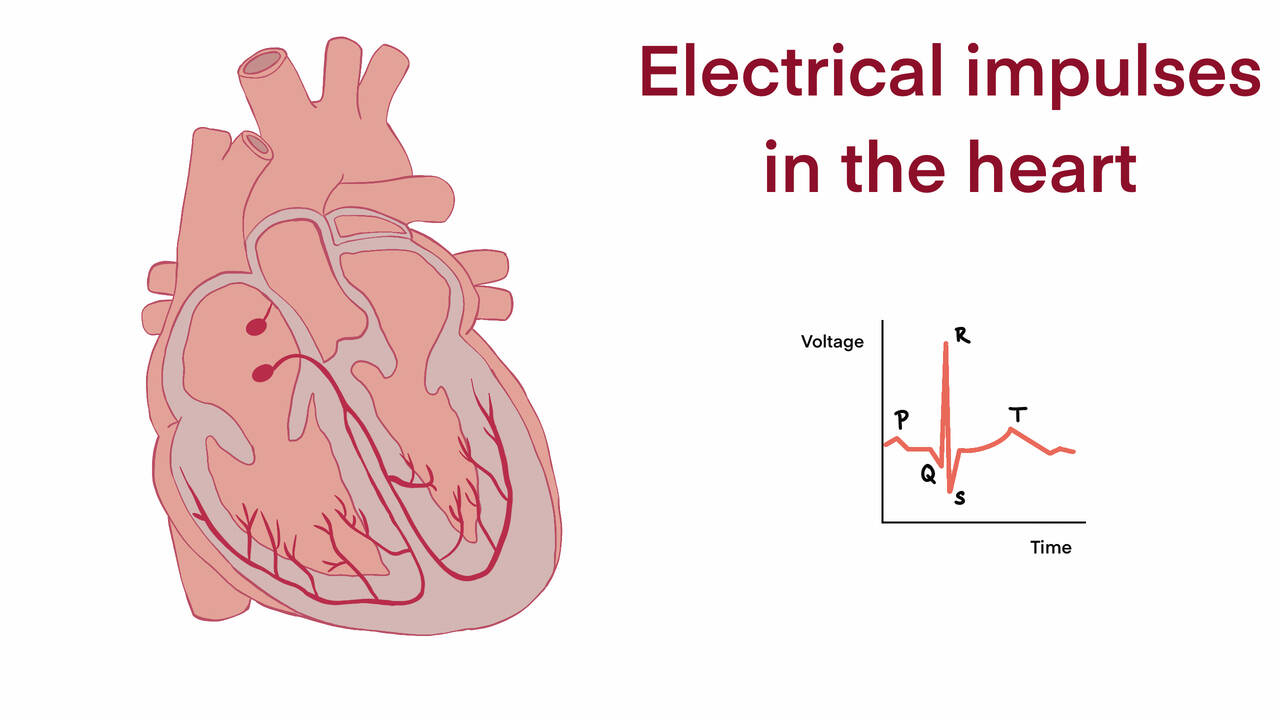
You’re starting a workout: music playing, you start to run. After a few minutes, it begins to feel like work and your heart starts pumping faster and faster. Just like any other muscle in your body, your heart works by reacting to electrical signals. It’s those signals that are making your heart pump faster and allowing you to get a good workout.
Lesson Objectives:
In this lesson you will:
- Understand the parts of the heart, and how they allow your body to function properly.
- Learn how electrical signals can be interpreted to visualize the heart and its functions.
While relatively small, voltage is flowing throughout your body. These electric pulses—which are about 1 millivolt—are enough to tell your muscles to contract or relax. You can try this right now by taking your arm and flexing your bicep!
What you just did was due to electrical signals in your body! Since the heart is involuntarily controlled, you won't be able to flex it like with other muscles. Why would that be, though?
It makes sense, especially when we consider how much work the heart does in a day! In 24 hours, the heart will beat around 100,000 times and pump a total of 190,000 gallons of blood. If the heart was a voluntary muscle you'd better be good at multitasking!
The heart is made up of four chambers, known as the left and right atrium and the left and right ventricle. Each side works similarly. Blood comes into the heart on both sides through the atria. Then, the blood moves to the ventricles, where it is pushed out into the body. We'll explore how the heart pushes blood out in just a little. First, it might be helpful to understand the difference between the left and right sides of the heart.
The right side of the heart pumps blood that is unoxygenated. Blood that comes through the right side of the heart will be pumped to the lungs, where it is oxygenated. After becoming oxygenated in the lungs blood returns to the left side of the heart. The left side of the heart pumps blood that is oxygenated throughout the body—allowing it to function properly.

Those electrical signals that we talked about are what allow the heart to pump, or move blood. Inside of the right atrium is specialized tissue called the sinoatrial node. This tissue is what starts pumping the heart. Each cell in the atrium exposed to the voltage momentarily contracts and passes the charge onto neighboring cells. This continues and is known as a depolarization wave. When the signal reaches the atrioventricular node more specialized fibers pick up the pulse. These are known as Purkinje fibers and can be seen branching out in both ventricles. In the same way that the charge made the atria contract, both ventricles will contract. Finally, when the ventricles return to their normal state the sinoatrial node starts the process over again.

Since the heart uses electricity, we can use specialized tools called electrocardiographs to analyze and diagnose problems! Electrocardiographs measure the pulses of voltage and produce a graph, known as an electrocardiogram(EKG).

By looking at the EKG above we can learn a lot about how the heart works!
- At pulse P—the atria are contracting.
- At pulse QRS—the ventricles are contracting.
- At pulse T—the ventricles are returning to their normal state.
Healthcare professionals are trained to use tools such as the electrocardiogram in order to understand and diagnose problems and diseases in the heart.